Category:Brodmann area 17
Jump to navigation
Jump to search
| Brodmann areas | ||||||||||||||||||||||||||||||||||||||||||||||||||||||||||||
| ||||||||||||||||||||||||||||||||||||||||||||||||||||||||||||
Brodmann area 17 is Primary visual cortex.
See also
Subcategories
This category has the following 2 subcategories, out of 2 total.
M
P
- Pericalcarine cortex (1 F)
Media in category "Brodmann area 17"
The following 32 files are in this category, out of 32 total.
-
 BA17 Primary visual cortex - animation.gif 300 × 300; 1.77 MB
-
 BA17 Primary visual cortex - lateral view.png 1,200 × 1,200; 689 KB
-
 BA17 Primary visual cortex - medial view.png 1,200 × 1,200; 532 KB
-
 BA17 Primary visual cortex - posterior view.png 1,200 × 1,200; 436 KB
-
 BA17,18,19 - Visual cortex (V1, V2, V3) - animation.gif 300 × 300; 1.62 MB
-
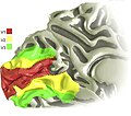
 BA17,18,19 - Visual cortex (V1, V2, V3) - lateral view.png 1,200 × 1,200; 693 KB
-
 BA17,18,19 - Visual cortex (V1, V2, V3) - medial view.png 1,200 × 1,200; 533 KB
-
 BA17,18,19 - Visual cortex (V1, V2, V3) - posterior view.png 1,200 × 1,200; 441 KB
-
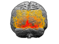
 Brain image of blind echolocator.tif 1,200 × 923; 882 KB
-

-
 Brodmann area 17 in the human brain (K. Brodmann, 1909, p. 217, Fig. 124-125).jpg 1,806 × 1,111; 355 KB
-
 Brodmann areas 17 18 19.png 256 × 192; 40 KB
-
 Brodmann Cytoarchitectonics 17.png 599 × 952; 231 KB
-
 Cortex strie.svg 305 × 393; 39 KB
-
 Ferret brain.jpg 850 × 505; 137 KB
-
 Mouse and human visual systems.jpg 850 × 731; 148 KB
-
 Occipital lobe in orangutan (K. Brodmann, 1909, p. 221, Fig. 136-137).jpg 1,402 × 1,098; 287 KB
-
 Parallel motion signals to V6 and MT+.jpg 397 × 548; 160 KB
-
 Pinwheel orientation.jpg 850 × 659; 66 KB
-

-
 Precentral and postcentral gyri in human (K. Brodmann, 1909, p. 153, Fig. 94-95).jpg 1,697 × 2,439; 959 KB
-
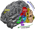 Presumed brain network processing the age of faces.png 1,243 × 1,022; 1.17 MB
-
 ProjectionsV1-V2.svg 393 × 283; 28 KB
-

-

-
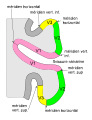 Scissure calca1.svg 354 × 461; 45 KB
-
-
The-Influence-of-Mexican-Hat-Recurrent-Connectivity-on-Noise-Correlations-and-Stimulus-Encoding-Video2.ogv 1 min 34 s, 2,000 × 2,000; 58.71 MB
-
 Thickness of an humn adult cerebral cortex.jpg 926 × 284; 148 KB
-
 V1 v2 v3.jpg 522 × 460; 32 KB
-
 Visual cortex.jpg 589 × 432; 37 KB
-
 Visualcortex.gif 201 × 179; 10 KB


_-_animation.gif)
_-_lateral_view.png)
_-_medial_view.png)
_-_posterior_view.png)
.jpg)


.jpg)


.jpg)
.jpg)


.jpg)